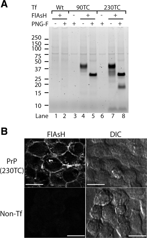
Figure 1.
IDEAL-labeling of cell surface TC-PrP with FlAsH. (A) Fluorescent gel analysis of cell lysates from IDEAL-labeled cells labeled with FlAsH. Cells were transfected with either wild-type PrP (lanes 1 and 2) or the indicated TC-PrP (lanes 3–8) expression construct. Samples where FlAsH was omitted show autofluorescent proteins (lanes 3 and 6). PNG-F indicates samples deglycosylated with PNGase F. (B) Confocal microscopy images of IDEAL-labeled cells labeled with FlAsH. PrP(230TC), cells expressing PrP with TC motif at position 230. Non-Tf, nontransfected control cells. DIC, differential interference contrast. Bars = 20 μm.

