
Figure 3.
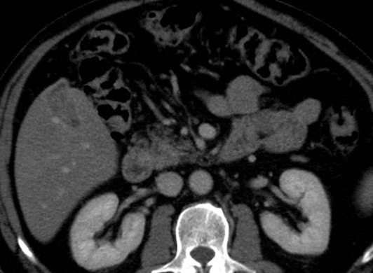
Transverse CT scan. It reveals a solid focal mass, about 3 cm in diameter, hypodense respect to the surrounding tissue in the basal phase (1) with a light peripherical enhanced rim in the arterial phase (2) that increases in the portal venous phase (3). In late phase (4), the lesion shows a puntacte aspect because of the presence of intralesional hypervascular spots (arrow). The bile ducts are not dilatated.