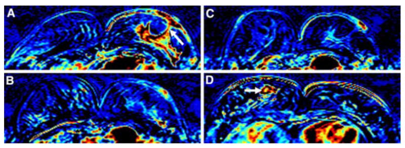
Figure 2.

A 47 year-old woman with IBC. (A) Baseline breast MRI prior to the NAC shows a big lobulated enhanced tumor in the left breast with skin invasion through the nipple (arrow). (B) After 1 cycle of AC, both the primary tumor and the skin enhancement subsided remarkably. (C) After 4 cycles of AC, the tumor response is even more obvious compared to the image after 2 cycles of AC. (D) After completion of NAC, combining AC plus TCH, both the main tumor and the skin lesion disappear. CCR was hence diagnosed. Note an enhanced lesion, diagnosed as mastitis, in the right breast (arrow). Final pathology after surgery, however, showed small islands of residual invasive cancer cells distributed in an area of 4.7cm in the left breast.
