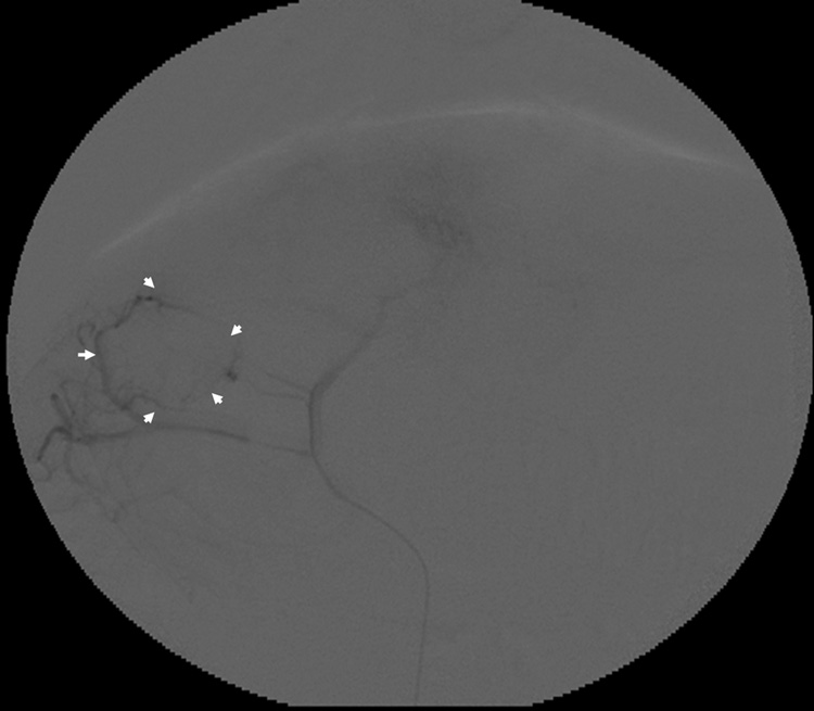
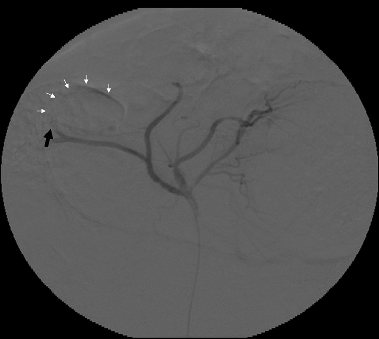
Figure 3.
Representative rabbit hepatic arteriograms before (a) and after (b) embolization of the left hepatic artery, which supplied the targeted VX2 liver tumor. The peripheral portion of the tumor is hypervascular (white arrows) prior to embolization (a). After embolization (b) there is abrupt cut off (black arrow) of the feeding artery without any remaining peripheral hypervascularity.


