
Figure 1. Colocalization of gB and CD63 in HHV-6-infected and uninfected HSB-2 cells.
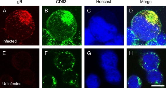
A–D) HSB-2 cells infected with HHV-6A by cell-to-cell contact and fixed at 4-day post-infection. E–H) Uninfected cells. The cells were stained with antibodies against gB (A and E) and CD63 (B and F) and with Hoechst 33258 (C and G). The merged panel shows the colocalization of gB with CD63 (D or H). Single sections were shown in this study. Scale bar: 10 μm.