
Figure 2. Morphological features of HHV-6-infected HSB-2 cells.
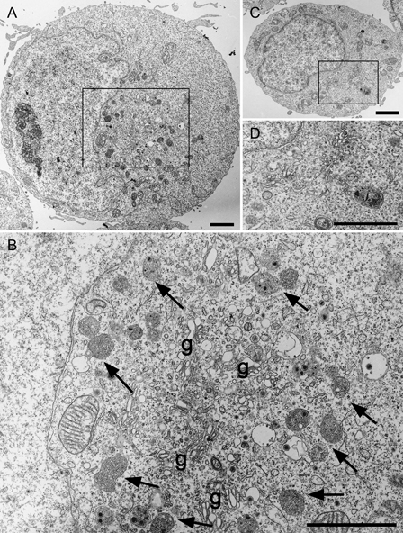
Ultrathin sections of Epon-embedded cells. Low (A and C) and high [(B) is boxed area in (A) and (D) is boxed area in (C)] power views of HHV6-infected (A and B) and uninfected (C and D) cells. Arrows indicate MVB-like membrane structures around several profiles of the Golgi apparatus (g). Scale bars: 2 μm.