
Figure 8. Accumulation of gB, gM and CD63 in MVBs.
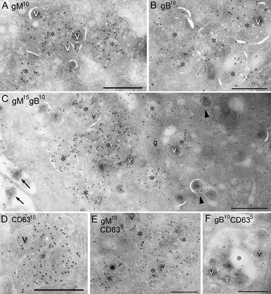
Immunogold labeling on ultrathin cryosections of HHV6A-infected cells showing gM (A) (10 nm), gB (B) or CD63 (D) (10 nm) or double labeling indicating gM (15 nm) and gB (10 nm) (C) or CD63 (5 nm) and gM (10 nm) (E) or gB (10 nm) (F). A–C) Immunogold particles indicating gM and/or gB were localized to both the internal vesicles of MVBs (asterisks) and the virions (v) within them. Note that extracellular virions (arrows), smaller vacuoles that contained only virions (arrowheads) and the Golgi apparatus (g) were also immunopositive for gM and gB. D) CD63 was localized to the internal vesicles of the MVBs (asterisk) and the virions (v) within them. E and F) Immunogold particles indicating gM (E) or gB (F) with CD63 were colocalized to the internal vesicles of the MVBs (asterisks) and the virions (v) within them. Scale bars: 0.5 μm.