
Figure 9. Exosomes contribute to the release of enveloped virions.
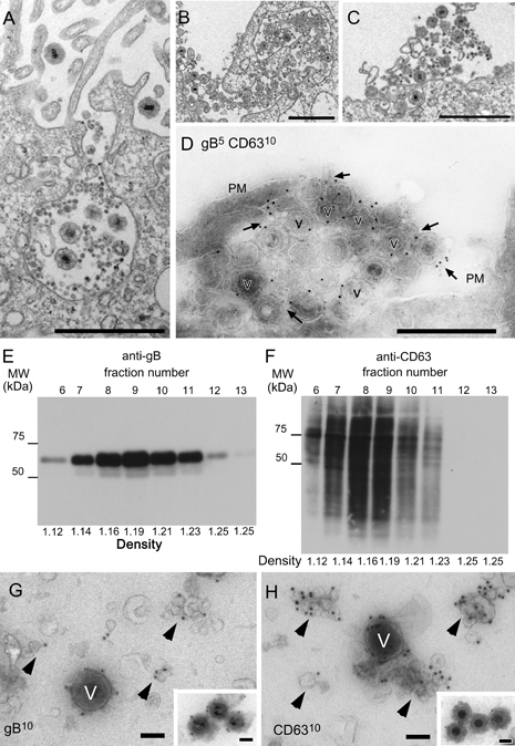
A–C) Ultrathin sections of Epon-embedded cells. MVBs were exocytosed from the cell surface, and numerous virions along with small internal vesicles were released from the cells. Released virions and exosome-like small internal vesicles were detected in the vicinity of the cell membrane (B and C). D) Double immunogold labeling of gB (5 nm) and CD63 (10 nm) in HHV6A-infected cells on ultrathin cryosections. Some virions (v) and exosomes (arrows) were positive for both gB and CD63. PM, plasma membrane. E–H) Exosome fractions containing virions were collected by sucrose density gradient from the culture medium of HHV-6A-infected T cells and were analyzed by immunoblotting and EM. E and F) Immunoblot analysis with anti-gB (E) or anti-CD63 (F) antibodies of the sucrose density gradient fractions. The same volume of the gradient fraction was used for all blots. Densities of the fractions are listed at the bottom in grams/milliliters. G and H) Immunogold labeling of gB (G) and CD63 (H) in whole-mount virions and exosomes from fraction 9 in panels (E) or (F). Gold particles showing gB (G) and CD63 (H) labeling of exosomes (arrowheads) and virions (v). Insets in (G and H) showed immunogold labeling of gB and CD63 in purified virions, respectively. Gold particles indicating gB [an inset in (G)] and CD63 [an inset in (H)] were detected in purified virions (v). Scale bars: 1 μm (A–C), 0.5 μm (D), 100 nm [(G and H) and insets in (G and H)].