
Figure 10. The infectivity of viruses secreted through the exosomal release pathway.
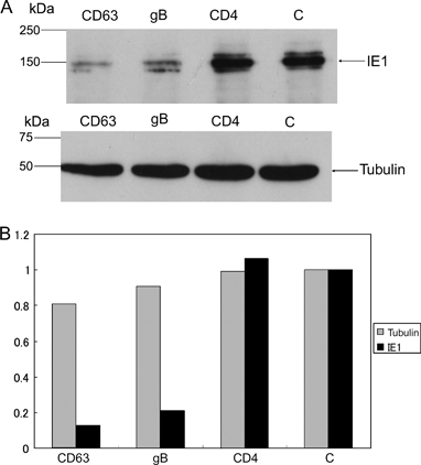
The supernatants of the medium of HHV-6-infected cells were collected and incubated with mAb against CD63 (CD63), gB (gB) or CD4 (CD4) at 4°C for 16 h. The supernatants were also incubated without mAb (C). Immunoprecipitation of viruses was performed by using mAb-based virus precipitation assay as described in Materials and Methods. The viruses non-immunoprecipitated were incubated with HSB-2 cells at 37°C for 1 h, and at 48-h post-incubation, the cells were harvested and lysed. A) Western blot of the lysates was performed with anti-IE1 or tubulin mAb. B) Quantitative analysis of western blot by kodak mi software shows the intensity of the band relative to that of C (without mAb). One of three independent experiments was shown. The mAb for CD4 was used as a negative control. The mAb for HHV-6 immediate early protein (IE1), which is not a viral structural protein, was used for the examination of HHV-6 infectivity, and the mAb for tubulin was used as an internal control of proteins.