
Figure 11. Intracellular distribution of the Golgi apparatus, MVBs and AL in an HHV6A-infected cell.
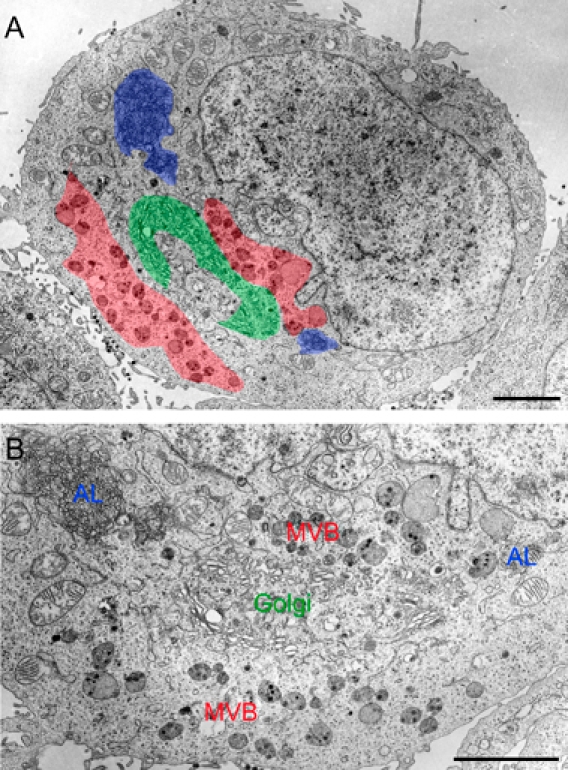
Ultrathin sections of Epon-embedded cells. A cluster of Golgi apparatus together with tubulo-vacuolar structures [marked by green color in (A) and Golgi in (B)] was located in the center of surrounding MVBs [marked by red color in (A) and MVB in (B)]. AL [blue in (A) and marked ‘AL’ in (B)] were observed apart from the Golgi apparatus and MVBs. Scale bars: 3 μm.