Fig. 3.

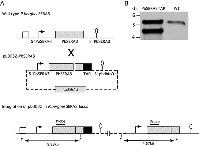
Integration of a TAP-tagged copy of PbSERA3 into the SERA locus. Shown are schematic representations detailing the outcome of the expected integration event.
A.Wild-type PbSERA locus showing part of the PbSERA2 gene (white box) and the PbSERA3 gene (grey box) with 5′ UTR and 3′ UTR (upper part). The pL0032-PbSERA3 targeting construct including the 5′ UTR and full-length PbSERA3 gene (without STOP codon) fused to a TAP-tag (black box), the 3′ pbdhfr/ts sequence and a selectable tgdhfr/ts marker cassette (middle part) was integrated into the PbSERA3 locus (lower part). F = FnuI restriction sites. The expected fragments are indicated.
B.Southern blot analysis showing the correct integration event. Wild-type parasites and transfected parasites were isolated from infected mice and genomic DNA was prepared. Upon FnuI digestion, Southern blot analysis was performed using a PbSERA3-specific DNA probe. The location of the probe is indicated in (A).
