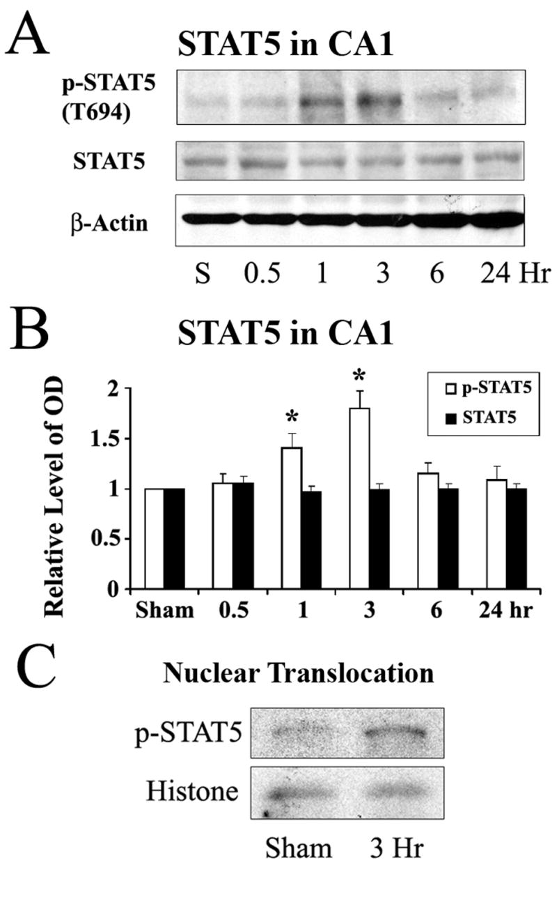
Figure 1. Increased phosphorylation of STAT5 in rat hippocampal CA1 in the early stage following global cerebral ischemia.

(A) Representative Western blots showing the levels of phosphorylated and total STAT5 in rat hippocampal CA1 at serial time points following ischemia. (B) The average levels of p-STAT5 in the CA1 region determined by Western blot were increased at 1 hr and 3 hr following ischemia compared to the sham-operated group. Data are mean ± SEM, assessed by ANOVA and post hoc Scheffe’s tests. *P<0.05 vs. the sham-operated. (C) Representative Western blot with nuclear preparation of hippocampal CA1 demonstrating increased nuclear translocation of p-STAT5 at 3 hr after ischemia.
