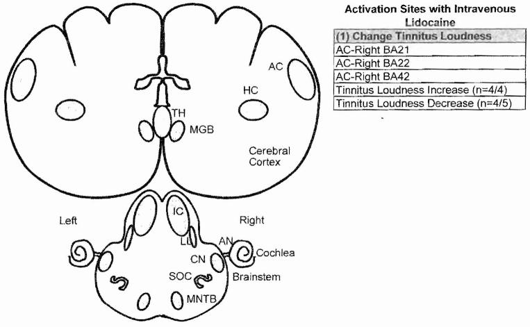
Figure 2.
Regions of auditory pathway that showed a significant change in activity (regional cerebral blood flow) measured with positron emission tomography during intravenous lidocaine treatment. Intravenous lidocaine increased tinnitus loudness in four patients; this was associated with increased activity in the right auditory cortex (AC). In four of five subjects, intravenous lidocaine decreased tinnitus; this was associated with decreased neural activity in the right AC. AN, auditory nerve; BA, Brodmann’s area; CN, cochlear nucleus; HC, hippocampus; IC, inferior colliculus; LL, lateral lemniscus; MGB, medial geniculate body; MNTB, medial nucleus of trapezoid body; SOC, superior olivary complex; TH, thalamus

