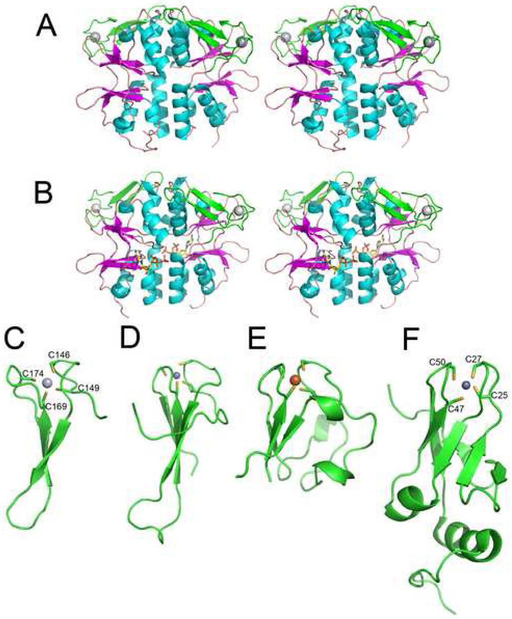
Figure 7.
Structures of TA0289 and other Zn ribbon domain proteins. (A), overall structure of the TA0289 dimer (stereo view). CBS domains are shown in orange (β-strands) and cyan (α-helices), Zn ribbon domains are shown in green, and metal atoms are shown as grey spheres. (B), stereo view of the TA0289 dimer shown with docked ATP from in silico analysis. (C), structure of the TA0289 Zn ribbon-like domain showing the tetrahedral coordination of the metal atom (Hg2+) by conserved cysteines (labeled). (D), structure of the Zn ribbon domain of the human transcriptional elongation factor TFIIS (1tfi). (E), structure of the P. furiosus rubredoxin PF1282 (1brf). (F), structure of KTI11 (1yop, 1yws). Metals are shown as: dark gray spheres (zinc; D and F), light gray sphere (mercury; C), and orange sphere (iron; E).

