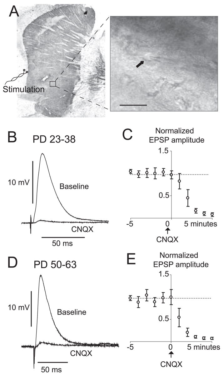
Fig. 2.
EPSPs evoked by electrical stimulation of the white matter adjacent to the NA and carrying cortical fibers in preadolescent and adult rats. (A) Tyrosine hydroxylase-stained slice illustrating the position of recording and stimulating electrodes. Inset shows an IR-DIC image of a MSN in the NA (arrow). (B) Representative corticoaccumbens EPSP in a slice from a preadolescent rat showing an overlay of EPSP evoked before (baseline) and after CNQX (10 μM). Traces are average of five repetitions. (C) Population data of EPSP amplitude normalized to baseline levels. Values are averages per minute. (D) Overlay of representative corticoaccumbens EPSPs in a slice from an adult rat showing blockade with CNQX. (E) Time course of normalized EPSP amplitudes for all the neurons tested. Calibration bar, 25 μm.

