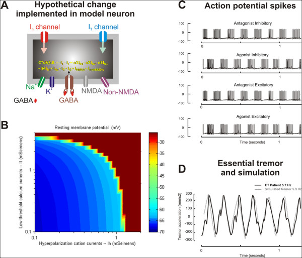
Figure 2.
(A) A traditional Hodgkin-Huxley model of cell membranes with multiple ion channels was used to generate the action potential. In order to simulate physiologically realistic neural behavior, ion channels such as hyperpolarization activated cation currents (Ih) and low threshold calcium current (IT) were also included. NMDA and non-NMDA excitatory glutamatergic channels as well as GABA sensitive inhibitory channels were also included. The grey box schematizes the burst neuron, while its grey outline schematizes the cell membrane. The ion channels span the membrane thickness. dV is the rate of change in the membrane potential over period 'dt'. C is the membrane capacitance (1 μF/cm2) and n1-n4 is a rate scaling factor determining the ion channel expression profile in the neuron. IL, IT, Ih1-4, INa, and IK, denote the leak current, low-threshold calcium current, hyperpolarization activated current (carried by HCN1-4), fast sodium current and delayed rectifier potassium current, respectively. ICl is the synaptic current mediated by glycinergic and GABAergic neurotransmitters. INMDA and InonNMDA are synaptic currents mediated by NMDA and non-NMDA sensitive glutamate receptors. (B) The effects of changing Ih (x-axis) and IT (y-axis) on the resting membrane potential (color coded) in the simulated neuron. As expected, increases in Ih and IT further depolarize the neuron. A depolarizing shift in the resting membrane potential reflects increased neural excitability. (C) Illustration of bursts of action potential spikes from the agonist and antagonist burst neurons. The alternate spiking behavior of these neurons is evident when they are plotted along the same time scale (x-axis). (D) Simulation (grey trace) of essential tremor (black trace) is shown. The tremor amplitude (y-axis) is plotted against time (x-axis). The time scale for simulated essential tremor is the same as the time scale for the traces representing the spiking behavior of the burst neurons. The frequency of tremor recorded from the patient is 5.7 Hz, which is closely simulated by the neuromimetic model (5.9 Hz). The amplitude of the simulated tremor also resembles the one recorded from the ET patient.

