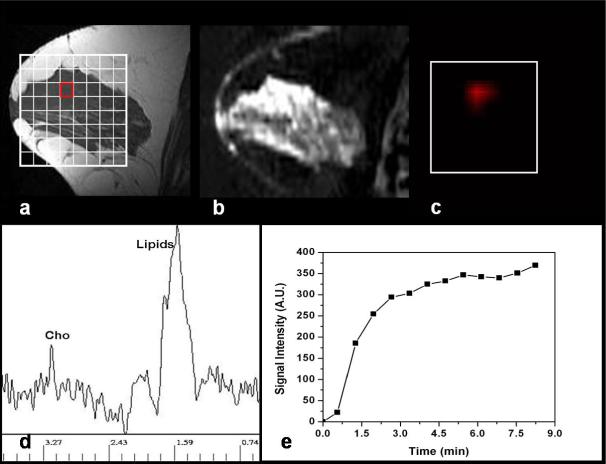
Figure 3.
MR imaging and chemical-shift imaging from a 45-year-old patient (#33) with benign fibroadenoma. (a) The sagittal T1-weighted pre-contrast MR image shows very dense glandular tissues in the right breast. (b) The sagittal view enhancement map shows a heterogeneously enhanced area in the anterior right breast. (c) The Cho metabolite map demonstrates elevated signal in the enhanced lesion. (d) MR spectrum from a voxel (red outline) clearly demonstrates a Cho peak, with Cho SNR = 6.5. (e) The signal enhancement time course from tissues corresponding to the Cho voxel shows the benign pattern with persistent enhancements during the delayed phase.

