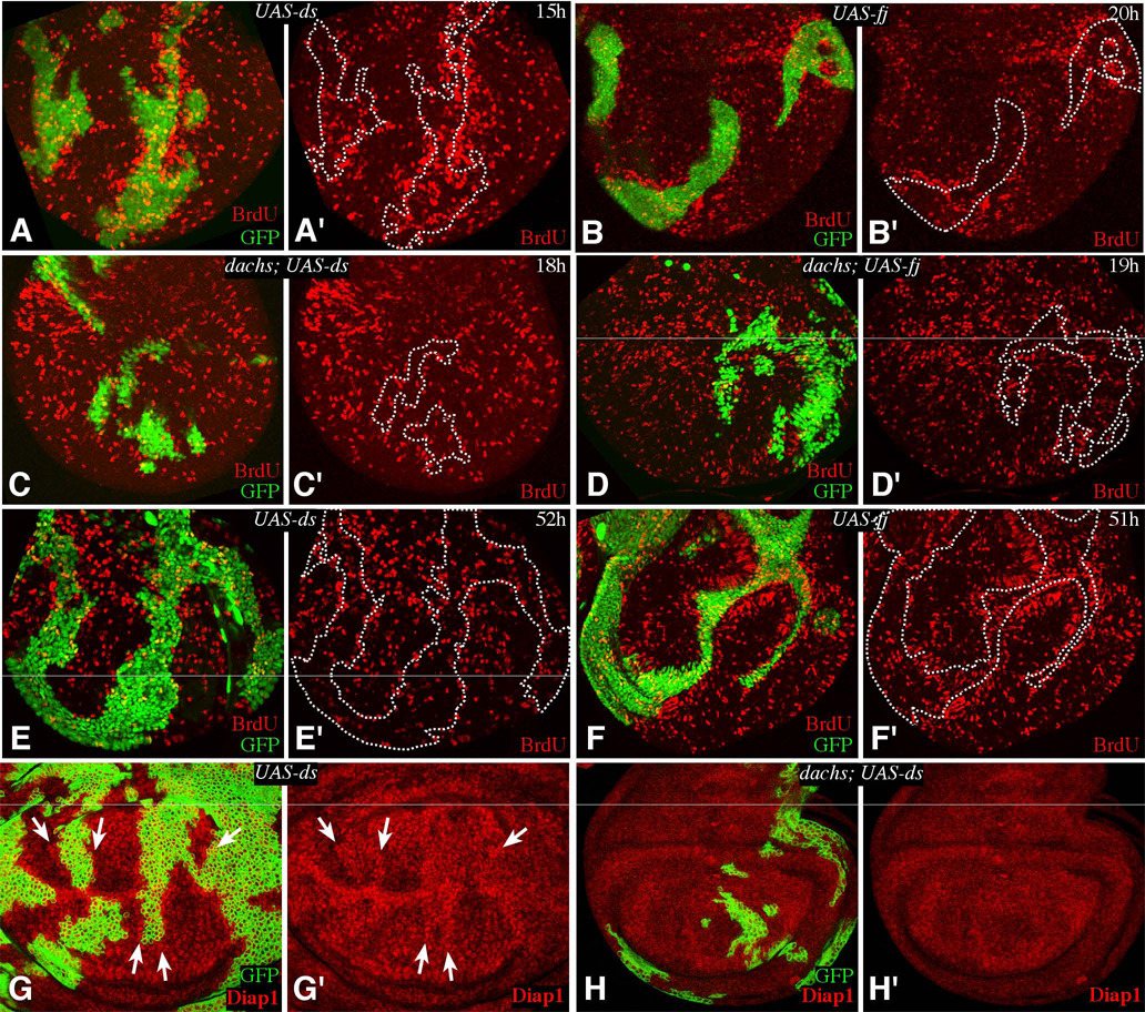
Figure 5. Fj- or Ds-expressing clones elevate BrdU incorporation.
A–F show wing imaginal discs containing Gal4:PR-expressing clones, marked by expression of GFP (green), grown for the indicated number of hours on media containing RU486, and labeled and stained for BrdU. For ease of comparison, the locations of selected clones are outlined by dashes. A,E) AyGal4:PR UAS-ds UAS-GFP. Elevated BrdU labeling is evident in A, especially in distal regions, but not in E. B,F) AyGal4:PR UAS-fj UAS-GFP. Elevated BrdU labeling is evident, especially in proximal regions. C) dachsGC13; AyGal4:PR UAS-ds UAS-GFP. BrdU labeling is not affected by the clones. D) dachsGC13; AyGal4:PR UAS-fj UAS-GFP. BrdU labeling is not affected by the clones. G) tub-Gal4/Gal80ts clones expressing ds; Diap1 staining is elevated around the clones (arrows). H) dachsGC13 mutant with tub-Gal4/Gal80ts clones expressing ds, Diap1 staining is not affected by the clones.

