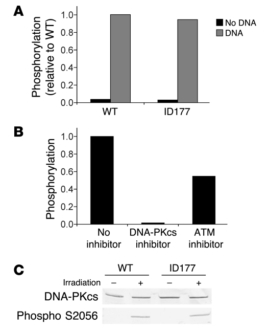
Figure 4. Measurement of DNA-PKcs activity.
(A) DNA-PKcs kinase activity of cellular extracts of wild-type (MRC5) and ID177 fibroblasts was measured by quantification of phosphorylation of a p53 peptide in the presence and absence of DNA. (B) DNA-PKcs kinase activity in ID177 cellular extracts was measured by quantification of phosphorylation of a p53 peptide in the presence of DNA without inhibitor or with DNA-PKcs–specific inhibitor NU7441 (33) or ATM-specific inhibitor KU-55933 (34) in a final concentration of 0.5 μM. Phosphorylation is expressed relative to ID177 without inhibitor. (C) Western blot analysis of DNA-PKcs and phosphorylated DNA-PKcs in cellular extracts from untreated (–) and irradiated (2 Gy) fibroblasts (+) of ID177 and wild-type (C5RO) fibroblasts with the DNA-PKcs antibodies 2,208 and 2,129 (1:1) and the phosphospecific DNA-PKcs antibody S2056.

