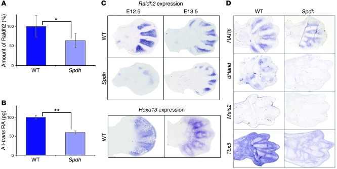
Figure 2. Dysregulation of RA pathway in Spdh.
(A) 2D gel electrophoresis of limb bud tissue (stage E13.5) demonstrated a reduction (63% of WT) of Raldh2 protein levels in Spdh mice (*P < 0.05). (B) Quantification of RA in E13.5 autopod limb tissue. Spdh limbs show a reduction to 57% when compared to WT (**P ≤ 0.005). (C) In situ hybridization against Raldh2 on forelimbs at E12.5 and E13.5 demonstrated a reduction of Raldh2 mRNA in Spdh limbs. In the WT limbs, Raldh2 was expressed in the interdigital space but not the cartilaginous condensations. At E13.5, Raldh2 expression was mainly found in the perichondrium. Expression overlapped with Hoxd13. (D) In situ hybridizations of WT and Spdh forelimbs at E13.5 of RA downstream targets RARβ, dHand, Meis2, and Tbx5. All showed reduced expression in Spdh limbs.

