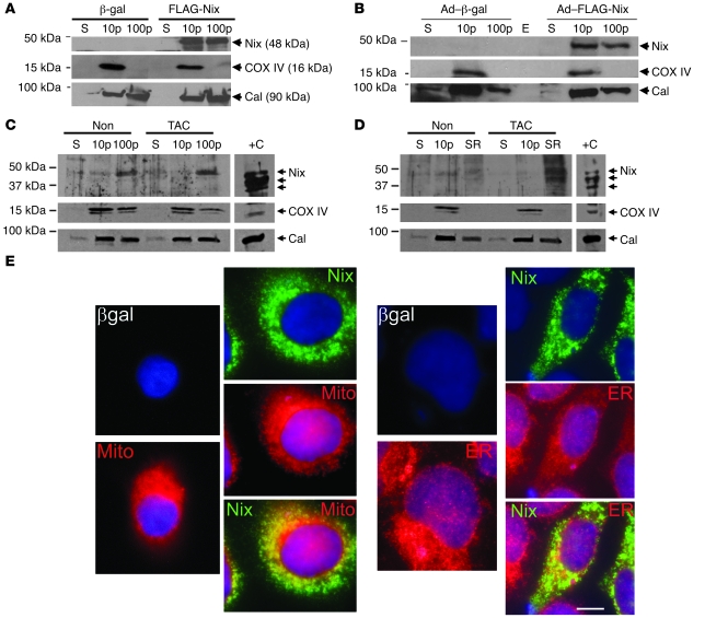
Figure 1. Nix localizes to mitochondria and ER and cofractionates with mitochondrial and ER/SR proteins.
(A) HEK293 cells were transfected with FLAG-Nix or β-gal control; fractionated into 10,000 g pellet (10p), 100,000 g pellet (100p), and 100,000 g supernatant (S); and immunoblotted with anti-FLAG, calnexin (Cal), or COX IV antibodies. (B) Neonatal rat ventricular myocytes were infected with adenoviruses encoding FLAG-Nix or β-gal control and processed as in A. E, empty lane. (C and D) Hearts from mice subjected to 1 week of pressure overload (TAC) and nonoperated controls (Non) were fractionated into a 10,000 g pellet and a 100,000 g pellet. The 100,000 g pellet was separated on a discontinuous sucrose gradient to yield the SR-rich fraction (SR, see Methods). 50 μg (C) and 20 μg (D) of the indicated fractions were separated by SDS-PAGE and immunoblotted with anti-Bnip3L (Nix), calnexin, or COX IV antibodies. Positive control (+C) (10 μg) was cellular extract from FLAG-Nix transfected HEK293 cells, showing multiple bands corresponding to Nix homodimers and heterodimers. (E) FLAG epitope–tagged Nix or β-gal control (both green) were transiently expressed in HEK293 cells and analyzed by fluorescence microscopy for colocalization with mitochondrial MitoFluor Red 589 (Mito) or ER calnexin (both red). Nuclei are blue (DAPI). Overlay for Nix is at bottom. Original magnification, ×1,000. Scale bar: 10 μm (shown for comparison).

