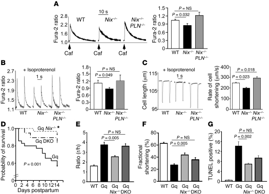
Figure 3. In vivo restoration of SR calcium stores in Nix-knockout cardiac myocytes by SERCA disinhibition reverses the Nix-null rescue of Gq peripartum cardiomyopathy.
(A) Ventricular cardiac myocytes isolated from WT, Nix-knockout (Nix–/–), or Nix/PLN-DKO (Nix–/–PLN–/–) mouse hearts were loaded with Fura-2 AM and analyzed for caffeine-stimulated [Ca2+]i. A representative set of tracings is shown (left). Group data (right) represent mean ± SEM of 35 WT, 28 Nix–/–, and 38 Nix–/–PLN–/– cardiac myocytes from n = 3 to 4 hearts each. (B) Pacing-stimulated [Ca2+]i in ventricular myocytes from the same groups. Representative tracings are shown (left) and group data represented as mean ± SEM (right). (C) Contraction of paced ventricular myocytes from the same groups. Representative contraction tracings are shown as the absolute change in cell length over time (left). Quantitative analysis of the peak rate of change of cell shortening are represented as mean ± SEM (right). Horizontal bars indicate time. (D) Kaplan-Meier analysis of mouse survival in the peripartum period. Daily survival of Gq (n = 36), Gq Nix–/– (n = 30), and Gq Nix–/–PLN–DKO (Gq DKO, n = 19) dams was assessed after giving birth. log-rank statistic was employed to detect statistical significance. *P = 0.017 versus Gq by post-hoc test (Holms-Sidak). (E–G) Comparative analysis of left ventricular dilation (E, measured as the ratio of ventricular radius [r] to wall thickness [h]), contraction (F, measured as echocardiographic percentage of fractional shortening), and apoptosis (G, measured as the percentage of TUNEL-positive cardiac myocytes) for the same study groups (n = 6–13/group). WT is shown for comparison with normal. Statistical significance was determined by 1-way ANOVA and Tukey’s post-hoc testing.

