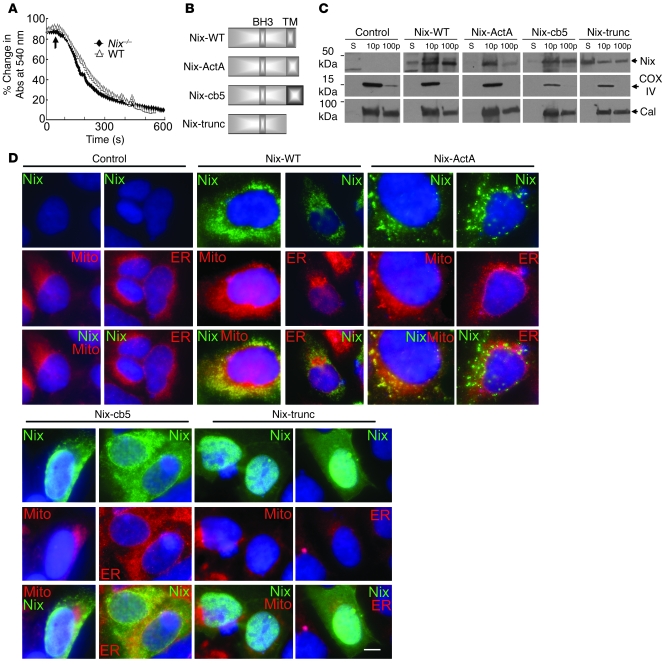
Figure 4. Creation of mitochondria- and ER-specific Nix mutants.
(A) Calcium-induced swelling of purified WT (triangles) and Nix-knockout (diamonds) liver mitochondria. Calcium (250 μM) was added (arrow) and the decrease in absorbance of 540 nm light assessed over time. Each curve represents the mean of 2 separate experiments. (B) Schematic depiction of mutation strategy for organelle-specific Nix. TM, putative transmembrane domain. (C and D) FLAG epitope–tagged WT Nix (Nix-WT), Nix-ActA, Nix-cb5, or truncated Nix (Nix-trunc) were transiently expressed in HEK293 cells, fractionated into 10,000 g pellet, 100,000 g pellet, and 100,000 g supernatant, and immunoblotted with anti-FLAG, calnexin, or COX IV antibodies (25 μg protein/lane). (D) Transiently transfected WT Nix, Nix-ActA, Nix-cb5, or truncated Nix (all green) as in C were analyzed by fluorescence microscopy for colocalization with MitoFluor Red 589 or ER calnexin (both red). Nuclei are blue (DAPI). Overlays are shown at bottom. Original magnification, ×1,000. Scale bar: 10 μm (shown for comparison).

