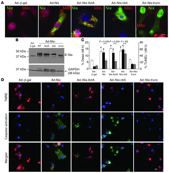
Figure 5. ER- and mitochondria-targeted Nix are equally effective in killing cultured cardiac myocytes but utilize different mediators.
(A) Cultured neonatal rat cardiac myocytes were infected with adenoviruses encoding WT Nix or 1 of the 3 Nix mutants and subjected to fluorescence microscopy for subcellular localization. Shown are overlay images with FLAG-Nix (green) and MitoFluor Red 589 (red). Original magnification, ×1,000. Scale bar: 10 μm (shown for comparison). (B) Cultured neonatal rat cardiac myocytes were infected with adenoviruses encoding WT Nix or 1 of the 3 Nix mutants, and Nix expression was analyzed by immunoblotting for FLAG epitope and GAPDH (loading control; 25 μg/lane). Arrows indicate Nix mutants with varying molecular weights. (C) Quantitative analysis of cardiomyocyte death (left y axis, white bars) and TUNEL positivity in the absence (right y axis, black bars) and presence (right y axis, gray bars) of 25 μM BAPTA-AM induced by subcellular targeting of Nix. Means ± SEM of 4 independent experiments for death and 8 (–BAPTA) and 5 for TUNEL (+BAPTA) are shown. (D) Confocal microscopy of TMRE (red) and fluorescent caspase substrate (rhodamine 100 bis-l-aspartic acid amide; green) in cultured neonatal rat cardiac myocytes infected with adenoviruses encoding WT Nix or 1 of the 3 Nix mutants. Original magnification, ×400. Nuclei are blue (Hoechst 33342). Scale bar: 20 μm (shown for comparison).

