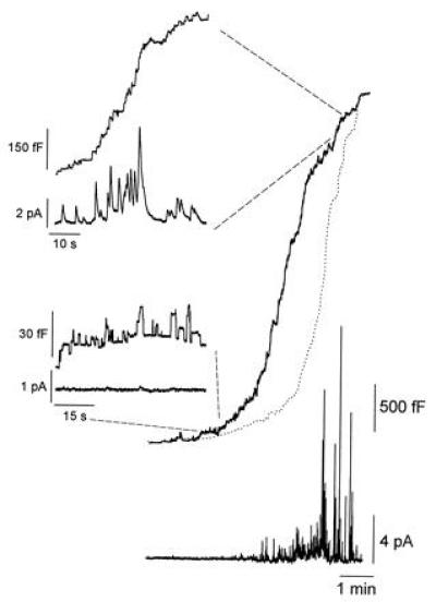
Figure 4.

In ruby-eye mouse mast cells, most of the fusion events seen during the initial phase of the degranulation show little or no release of serotonin. Simultaneous cell membrane capacitance and amperometric measurements of exocytosis in mast cells obtained from ruby-eye mice. The upper traces show the cell membrane capacitance increases during a complete degranulation. The lower traces show the corresponding spike like amperometric recordings of serotonin release. The time integral of the amperometric trace is shown as a dotted line superimposed on the cell membrane capacitance trace and represents the total amount of serotonin detected by the carbon fiber during the secretory response. Secretion was stimulated by adding 5 μM GTPγS to the patch-pipette solution.
