
Figure 15.
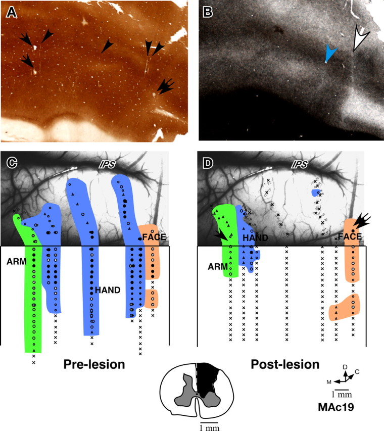
Somatotopy in area 3b of monkey MAc19 before and immediately after lesion of the dorsal columns. A, Cytochrome oxidase-stained section of the flattened cortex through area 3b showing electrolytic microlesions made during the experiment (arrows and double arrow) and some visible electrode tracks (arrowheads). B, A myelin-stained section of the flattened cortex showing the hand–face border (white arrow) and the border between D1 and D2 (blue arrowhead). C, Locations of neurons with receptive fields on the face, hand and arm encountered in area 3b and the adjacent cortex before lesion of the dorsal columns of the spinal cord, and (D) immediately after lesion of the contralateral dorsal columns of the spinal cord. Only a few neurons responded to the taps on the hand or arm perhaps because of sparing of some of the dorsal column fibers. The arrow and the double arrow shown in D indicate the penetrations marked with electrolytic lesions (compare with A). The photograph underlay in C and D shows the correspondence of the prelesion and postlesion penetration sites with respect to the surface vasculature of the brain. Intraparietal sulcus (IPS) is visible at the top of the photograph. Note that the hand–face border is located near the end of the IPS, our basis for interpretation of this border for monkey MCh21. Inset at the bottom shows the spinal cord lesion reconstructed from the transverse sections of the spinal cord. Conventions as for Figure 4 A.