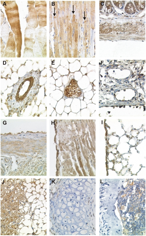
Figure 5. CK1δ expression in immobile cells of mesenchymal origin.
Fixative: acid formalin, fixation by immersion. Peroxidase reaction, dye: DAB. CK1δ specific antiserum: NC10. The CK1δ immunostaining results of striated muscle cells of the skeletal system (A), the myocardium (B), and of smooth muscle cells of the intestinal wall (C); aterial blood vessel (D); venous blood vessel (E); lymphatic vessel (F); mesothelial cells of the peritoneum, (G) mesothelial cells of the pericardium (H), mesothelial cells of the pleura (I), adipocytes of white and brown fatty tissue (J), chondrocytes of the hyaline cartilage (K) and osteocytes (L) are shown. In B, arrows indicate intercalated discs of cardiomyocytes. Magnification: 200× (G, H, J), 400× (A–F, I, K, L).

