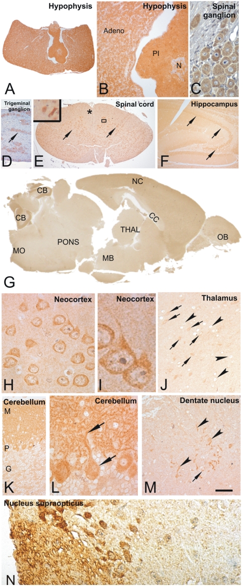
Figure 7. CK1δ expression in the nervous system.
Fixative: acid formalin, fixation by immersion. Peroxidase reaction, dye: DAB. (A) CK1δ was strongly expressed in the hypophysis. (B) The high-power view shows a cytoplasmatic staining of the epithelial cells of the adenohypophysis (Adeno). The pars intermedia (PI) and the neurohypophysis (N) were also strongly marked. (C, D): The nerve cell perikarya of a spinal (C) and a trigeminal ganglion (D, arrow) exhibit CK1δ whereas the adjacent nerve fibers were not marked. (E) The spinal cord neurons were strongly marked (arrows) whereas the gray matter neuropil and the white matter only faintly exhibited CK1δ (asterix). The inset demonstrates the high power view into the boxed area and shows a marked cytoplasmic labelling. (F) In the hippocampal formation the neurons of the sectors CA1, CA2 and CA3 were strongly labelled (arrows). (G) Sagittal section of a mouse brain immunostained with the NC10 antibody directed against CK1δ. There are especially high levels of CK1δ detectable in all layers of the neocortex (NC), the olfactory bulb (OB), and the molecular and Purkinje-cell layer of the cerebellum (CB). The white matter as seen in the corpus callosum (CC) and the cerebellar white matter did not show high levels of CK1δ. Low levels of CK1δ were found in the thalamus (THAL), midbrain (MB), pons (P) and the medulla oblongata (MO). (H, I) At higher magnification neocortical neurons show a strong cytoplasmatic staining whereas the neuropil was weakly stained. There was no staining of glial cells detectable. (J) In the thalamus there was a very light staining of the neuropil. Some thalamic neurons exhibited CK1δ in the cytoplasm (arrows) whereas other neurons were not labelled (arrowheads). (K) In the cerebellum CK1δ stained the molecular layer (M) and the Purkinje cell layer (P). The granule cell layer neurons (G) were not marked. There was only a very light staining of the neuropil in this layer. (L) At the higher magnification levels it was evident that the CK1δ expression in the molecular layer was the result of the staining of the entire dendritic trees of the Purkinje cells. The arrows indicate a Purkinje cell with the apical dendrite positive for CK1δ. There was no labelling of Bergmann glia cells. (M) In the dentate nucleus of the cerebellum the neuropil was weakly stained whereas some neurons were strongly CK1δ positive (arrows). Neurites were also labelled in this nucleus (arrowheads). Hypothalamic nuclei showed varying levels of CK1δ. Some of these neurons, e.g. in the suprachiasmatic nulceus exhibited nuclear CK1δ. N. supraopticus (N). Calibration bar in L equals: A = 270 µm, B = 120 µm, C, H, L, N = 180 µm, D = 150 µm, E = 400 µm, E-inset = 55 µm F = 280 µm, G = 800 µm, I = 6.6 µm, J, M = 40 µm, K = 70 µm. CK1δ was stained with the antibodies NC10 (A–C, E–N) and abcam 10877 (D).

