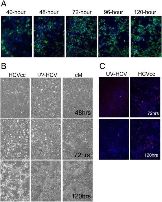
Figure 1. Characterization of HCV-JFH infection of Huh-7.5 cells.
(A) Expression of HCV NS5A in Huh-7.5 cells. HCV (+) cells are detected using an anti-NS5A antibody (green) while all cell nuclei are shown using Hoechst dye (blue). (B) Cytopathic effect observed during later time-points of HCV infection in Huh-7.5 cells. (C) Presence of activated caspase-3 (red) as shown by immuno-fluorescence staining of HCV-infected Huh-7.5 cells. Nuclei are shown using Hoechst dye (blue).

