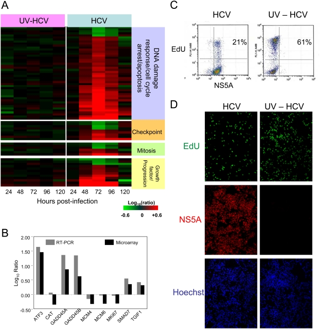
Figure 4. Cell cycle analysis during HCV infection.
(A) Gene annotation was performed using Ingenuity Pathway Analysis and Entrez gene. Expression profiles of sequences that are regulated (2-fold, P value<0.05) in at least 1 experiment. Each column represents gene expression data from an individual experiment comparing either UV-inactivated HCV or HCV-treated cells relative to time-matched, mock-treated Huh-7.5 cells. Genes shown in red were up-regulated, genes shown in green down-regulated, and genes in black indicate no change in expression in HCV-infected cells relative to uninfected cells. (B) RT-PCR validation of the expression array data. Data are shown as log10 ratio and reflect the difference in expression between HCV-infected (72 hours post-infection) and mock Huh-7.5 cells. (C,D) Analysis of proliferating cells at 72 hours post-infection with HCV or UV-inactivated virus (UV-HCV). Following a 3-hour pulse with the nucleoside analog EdU, cells were enumerated by flow cytometry (C) or visualized as adherent cells using immunofluorescence (D).

