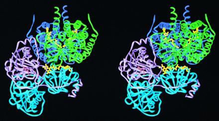
Figure 6.

A stereo ribbon diagram depicting the hypothetical docking of ETF to an MCAD dimer. The refined structures of human ETF and porcine MCAD (43) were used. The MCAD dimer (green and dark blue, mainly the green monomer) fits into the groove formed by the α (cyan) and β (lavender) subunits of ETF, in a manner close to the FAD of MCAD. By this mechanism, the closest flavin–flavin distance is approximately 19.5 Å.
