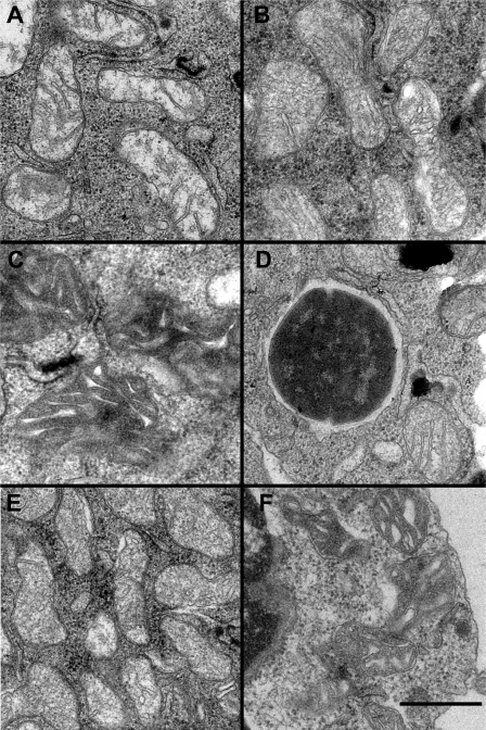
FIGURE 7.
Electron microscopy reveals cristae remodeling within mitochondria of GAS-infected cells. A, mitochondria in control cells treated with media display typical ultrastructure with the inner membrane projecting into the matrix at crista junctions to form lamellar cristae. B, mitochondria in GAS-infected cells show altered cristae structure consistent with swollen crista compartments. C, some mitochondria in GAS-treated cells have a different appearance with irregularly shaped cristae cross-sections within a very dense matrix compartment. D, Q-VD-OPH effectively prevents the alterations of mitochondrial structure found in GAS-infected macrophages. Several mitochondria, including the one at lower right, maintain normal crista structure despite being immediately adjacent to a phagocytosed GAS cell slightly left of center in this image. E, cells treated with purified SLO showing mitochondria with dilated cristae similar to those treated with GAS in B. F, mitochondria in SLO-treated cells with condensed matrix compartments similar to those following GAS treatment in C. Scale bar indicates 200 nm.

