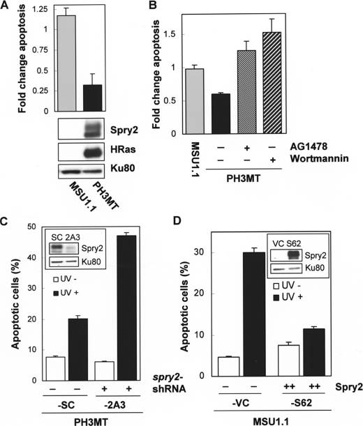
FIGURE 1.
Role of Spry2 on the inhibition of UV-induced apoptosis by HRas. A, normal parental non-transformed human fibroblasts (MSU1.1) and their HRas oncogene-transformed derivatives (PH3MT) were irradiated with UV (30 J/m2), incubated at 37 °C in media containing 10% serum for 4 h, and then assayed for early apoptotic events with annexin-FITC as indicated under “Experimental Procedures.” Cells that stained positive for annexin only were analyzed. The percentage of apoptotic cells following UV treatment was normalized to the percentage of cells undergoing apoptosis in the absence of treatment and is expressed in the graph as -fold change. A representative of three experiments is shown. A, MSU1.1 and PH3MT cells were treated and analyzed as described. A Western blot showing the level of Spry2 and HRas proteins is also shown. B, MSU1.1 and PH3MT cells were analyzed as described in the presence or absence of AG1478 (6 μm) and wortmannin (50 nm), as indicated. A representative of three experiments is shown. C, HRas-transformed cell lines with endogenous (PH3MT-SC) or down-regulated (PH3MT-2A3) levels of Spry2 (inset) were exposed to UV radiation (90 J/m2), and the percentage of cells undergoing apoptosis in the presence (black bars) or absence (white bars) of such treatment is shown. A representative of seven experiments is shown. D, the parental non-transformed human fibroblasts expressing an empty vector (MSU1.1-VC (VC)) or high levels of Spry2 (MSU1.1-S62 (S62)) (inset) were treated and analyzed as described. A representative of three experiments is shown.

