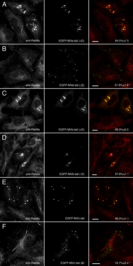
FIGURE 4.
Myosin Va, Vb, and Vc tails alter endogenous Rab8a distribution. A and B, HeLa cells transfected with EGFP-myosin Va tail +D or EGFP-myosin Va tail -D and stained for Rab8a. As with Rab10, EGFP-myosin Va tail +D caused endogenous Rab8a to mislocalize to EGFP-labeled puncta, whereas EGFP-myosin Va tail -D did not co-localize with Rab8a. C and D, expression of either splice variant of EGFP-myosin Vb tail (+Dor -D) caused Rab8a to redistribute to the EGFP-labeled perinuclear cisternum. E and F, only wild-type EGFP-myosin Vc tail, which contains an exon D-like domain, was able to recruit Rab8a, whereas MVc-tail-ΔD did not. Scale bars in all panels represent 10 μm. Percent co-localization (±S.E.) are listed in the merged images on the right of each panel (n ≥ 10). *, statistically significant difference comparing +D and -D constructs (p < 0.001).

