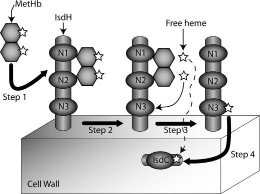
FIGURE 6.
Schematic showing how the IsdH protein may capture and transfer heme from hemoglobin. Step 1, MetHb is tethered to IsdH via the N-terminal IsdHN1 and IsdHN2 NEAT domains. MetHb is shown as two hexagons representing the α and β globin chains. Heme molecules are represented by five-pointed stars. Step 2, heme is released from MetHb. Our data indicates that this process is passive when NEAT domains are in isolation. Step 3, heme diffuses and is captured by the C-terminal IsdHN3 NEAT domain. Step 4, heme is released from IsdHN3 and subsequently captured by IsdC located within the cell wall. Our results also indicate that IsdC can directly capture heme that is passively released from MetHb (dashed line). Note that IsdH and IsdC are covalently attached to the cell wall by the SrtA and SrtB sortases, respectively.

