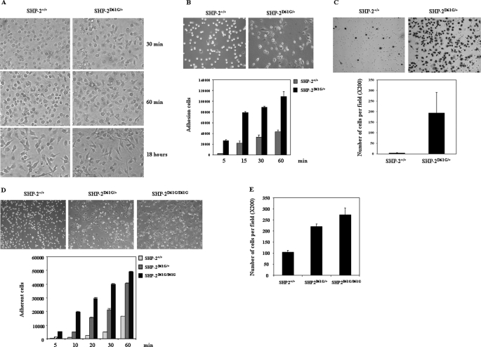
FIGURE 2.
Adhesion and migration of SHP-2 D61G knock-in cells are dramatically enhanced. A, WT and SHP-2D61G/+ bone marrow-derived macrophages were replated into non-tissue culture plates in cytokine-free medium and photographed 30 min, 60 min, and 18 h later. B, upper panel, WT and SHP-2D61G/+ bone marrow-derived mast cells were plated into cytokine-free medium and photographed 30 min later. Lower panel, WT and SHP-2D61G/+ mast cells were allowed to adhere to fibronectin-coated wells in cytokine and serum-free medium for the indicated periods of time. The number of adherent cells was determined by the MTS assay. C, cells were assayed for migration as described in the legend to Fig. 1C. Migrated cells adhering to the lower side of the membranes were photographed and enumerated under a microscope. D, immortalized WT, SHP-2D61G/+, and SHP-2D61G/D61G embryonic fibroblasts were suspended in DMEM with 2% BSA, allowed to adhere to fibronectin-coated plates for 6 h, and then photographed. Adhesion (D) and migration (E) capabilities of the cells were assessed as described under “Experimental Procedures.” Three independent experiments were performed and similar results were obtained in each. Results shown are the mean ± S.D. of triplicates from one experiment.

