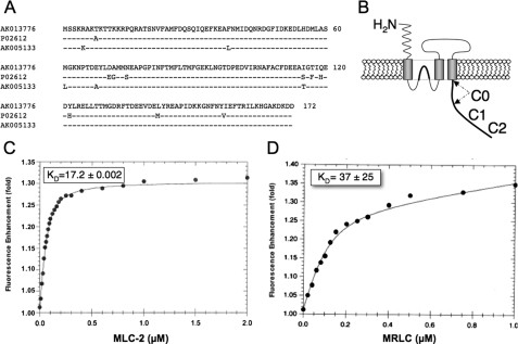
FIGURE 1.
Myosin RLC isoforms enhance fluorescence emission of a fluorescein-labeled peptide corresponding to the C0 region of the NMDAR1 carboxyl tail. A, amino acid sequence of a myosin RLC isoform previously identified in mouse hippocampus (GenBank™ accession number AK013776), the major smooth muscle RLC isoform in chicken gizzard (GenBank™ accession number P02612), and an RLC isoform identified from mouse cerebellum (GenBank™ accession number AK005133). AK013776 is identical to the isoform previously identified in mouse brain (MRLC) as an NMDA-receptor interacting protein (2). B, schematic representation of the NMDAR1 subunit showing the location of C0, C1, and C2 regions. C and D, titration of either purified smooth muscle MLC-2 (C) or purified, recombinant MRLC (D) enhanced fluorescence emission by ∼30%. Data were fit to a single hyperbolic curve using Equation 1 to derive binding affinities of 37 ± 25 nm (a = 1.0110 ± 0.0001, b = 2.6 ± 0.9, c = 0.09 ± 0.03, R0 = 0.10 ± 0.03, and r2 = 0.999) for MRLC (C), and 17.2 ± 2.2 nm (a = 0.996 ± 0.004, b = 7.42 ± 0.54, c = 0.029 ± 0.006, R0 = 0.073 ± 0.004, and r2 = 0.999) for MLC-2 (D). Each point represents fluorescence determined in quadruplicate; standard errors lie within the data points.

