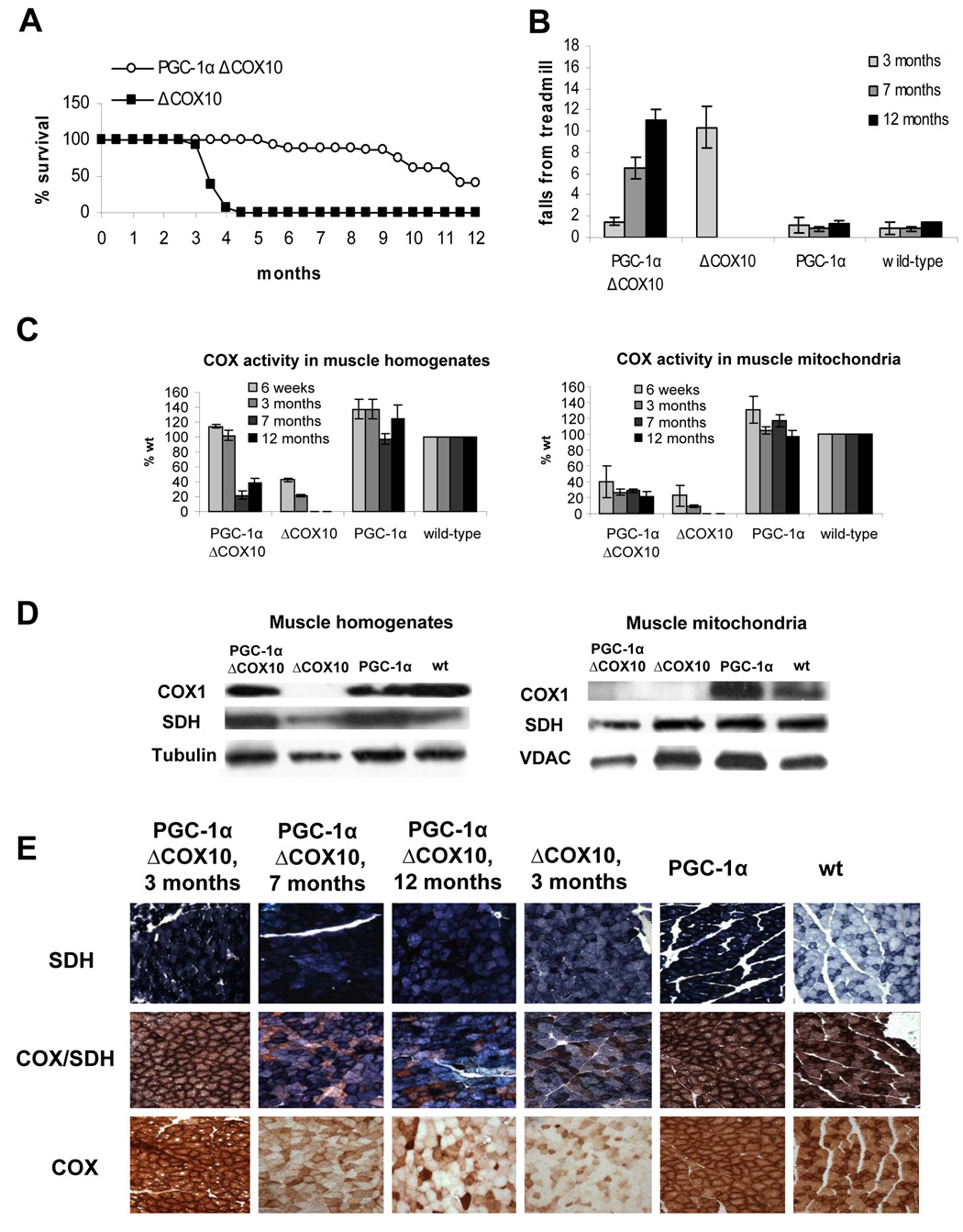
Figure 1. Longer life span and delayed onset of the myopathy coincide with a rescued COX activity muscle homogenates of PGC-1αΔCOX10 mice compared to ΔCOX10 mice.
A: Survival curve of female PGC-1αΔCOX10 mice in comparison to ΔCOX10 mice (n=20 for each group). No mice of the PGC-1α and wild-type control groups died in the observed timeframe. B: Treadmill performance test at different ages for female PGC-1αΔCOX10, ΔCOX10, PGC-1α and wild-type mice (n= 6 for each group). One way ANOVA, followed by Post-Hoc Tukey analyses showed significance between ΔCOX10 and each of the other groups. C: Cytochrome c oxidase (COX) activity of female PGC-1αΔCOX10, ΔCOX10, PGC-1α and wild-type mice at different ages comparing muscle homogenates and muscle mitochondria (n=3 for each group). D: Western blot of COXI, SDH and tubulin in muscle homogenates and mitochondria of 3 months old female PGC-1αΔCOX10, ΔCOX10, PGC-1α and wild-type mice. COXI levels were below the detection limit of the assay in some samples. E: Histology of the biceps femoris muscle from female mice at different ages (20x magnification) showing the development of the COX deficiency in PGC-1αΔCOX10 mice in comparison to a myopathic ΔCOX10 and to PGC-1α and wild-type mice as controls. Shown are succinate dehydrogenase (SDH), cytochrome c oxidase (COX) and combined COX/SDH staining.

