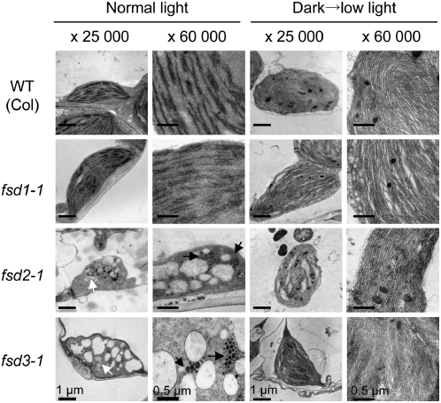
Figure 2.
Transmission Electron Micrographs of Plastids in fsd Mutants.
Leaves of wild-type (Columbia [Col] ecotype) and three FSD knockout mutant (fsd1-1, fsd2-1, and fsd3-1) plants were grown under a 16-h-day/8-h-night cycle for 10 d (left) or exposed to low light (photon flux density of 7 μmol·m−2·s−1) for 3 h after being grown in darkness for 10 d (right). Images were observed at low (×25,000) and high (×60,000) magnifications. White and black arrows indicate the abnormal suborganelle and the densely stained globular structures, respectively.

