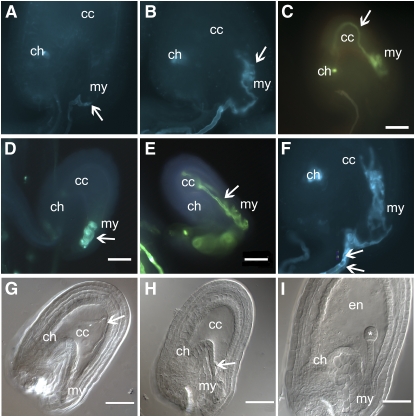
Figure 1.
Phenotype of lorelei Mutants.
(A) to (F) Aniline blue staining of ovules from different lorelei mutants pollinated with wild-type pollen.
(A) Wild-type arrival of the pollen tube (arrow) to the micropyle (my). There is no fluorescence inside the ovule.
(B) Pollen tube curling at the micropyle of an lre-1 ovule.
(C) Pollen tube invading the central cell (cc) and turning back toward the micropyle of an lre-2 ovule.
(D) Pollen tube curling inside the micropylar end of an lre-3 mutant.
(E) Pollen tube entering the central cell of an lre-4 ovule and heading back toward the micropyle.
(F) An example of two pollen tubes reaching the same lre-1 ovule. The two pollen tubes crawl along the funiculus and enter the embryo sac before forming coils at the micropylar end.
(G) to (I) DIC pictures of ovules the same lre-2/+ silique pollinated with wild-type pollen.
(G) An example of pollen tube entering deep inside the central cell and turning back toward the micropyle.
(H) Pollen tube coiling at the micropylar end of the embryo sac.
(I) Wild-type-looking ovule with a developed endosperm (en) and an embryo at the dermatogen stage (asterisk).
Arrow, pollen tube; cc, central cell; my, micropylar end; ch, chalazial end; en, endosperm; *, embryo. Bars = 50 μm.

