Figure 5.
Subcellular Localization of LORELEI.
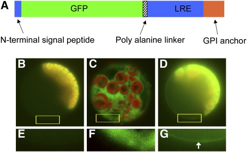
(A) Schematic representation of the GFP-LRE construct. The GFP is sandwiched between the N-terminal signal peptide and the rest of the LORELEI protein (central domain and GPI anchor domain). The predicted GPI anchor domain is shown in orange. An 8x Ala linker was added at the junction between the GFP and the major part of LORELEI.
(B) to (G) Epifluorescence detection of GFP-LRE in transfected protoplasts. The GFP fusion protein (green) appeared at the cell periphery as a line in transfected mesophyll protoplasts in which chloroplast autofluorescence (red) was also detected ([D]; detail from boxed region is shown in [G], with the arrow showing the thin fluorescent band around at the periphery of the cell). In protoplasts transfected with GFP only, the GFP protein (green) was seen throughout the cell, with no preference for the cell periphery. The autofluorescence from the chloroplasts appears in red ([C]; detail from boxed region is shown in [F]). In untransfected protoplasts, only chloroplast autofluorescence was detected (red) ([B]; detail from boxed region is shown in [E]). Exposure times for the pictures were as follows: 1 s for the green and red channels of the untransfected control ([B] and [E]), 0.6 s for the green and red channels of GFP ([C] and [F]), and 0.8 s for the green channel and 1 s for the red channel of GFP-LRE ([D] and [G]).

