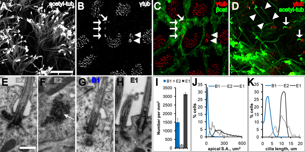
Figure 1. Three cell types contact the LV.
(A–D) Confocal images taken at the surface of wholemounts of the lateral wall of the LV.
(A) Acetylated-tubulin staining reveals many long cilia of ependymal cells. Scale bar = 10 um (A–D).
(B, C) γ-tubulin staining (B) together with β-catenin staining (C) reveals three cell types: E1 (many basal bodies), B1 (arrows indicate single basal body), and E2 (arrowheads indicate two basal bodies).
(D) Three cell types are also distinguished by their cilia. B1 cells have a single short cilium (arrows) and E2 cells have 2 long cilia (arrowheads).
(E, F) Electron micrographs from two serially reconstructed E2 cells. The central barrel of the basal body is cut transversely in (E) and in cross-section in (F) (arrow). Scale bar = 0.5 um (E–H).
(G) Electron micrograph of the primary cilium, basal body and centriole of a B1 cell. Note the simpler B1 basal body compared with the large, lobular E2 basal body (F). Both B1 and E2 (E) cilia were invaginated in the apical membrane.
(H) Electron micrograph of E1 cilia, which were not invaginated.
(I) Density of B1, E1, and E2 cells on the lateral wall of the LV. Error bars show sem.
(J) Histogram of apical surface area of B1, E1, and E2 cells. Data from 771 B1 cells, 1716 E1 cells, and 201 E2 cells from n=4 mice.
(K) Histogram of cilia length of B1, E1, and E2 cells. Data from 361 B1 primary cilia, 55 ependymal cilia, and 106 E2 cilia from n=3 mice.

