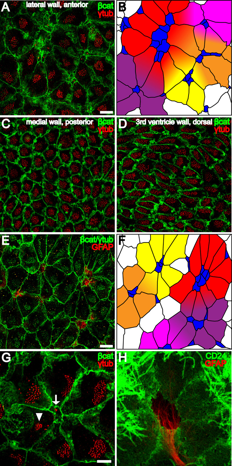
Figure 3. Pinwheel architecture of the ventricular surface in the adult neurogenic niche.
(A) Confocal image of the surface of the lateral wall of the LV stained for γ-tubulin (red) and β-catenin (green) reveals pinwheels with centrally located B1 apical surfaces surrounded by ependymal cells. Scale bar = 10 um (A–D).
(B) Tracing of the β-catenin+ membranes in (A) and color-coding of B1 cells (blue) and E1 cells (yellow, orange, red, purple and magenta) to illustrate 5 pinwheels. Some E1 cells occupy positions in more than one pinwheel.
(C,D) Neither B1 apical surfaces nor pinwheels are observed in the posterior medial wall of the LV or in 3rd ventricle walls.
(E) Confocal image of the surface of the lateral wall of the LV shows that pinwheels surround GFAP+ (red) B1 cells. β-catenin and γ-tubulin are in green. Scale bar = 10 um.
(F) Color-coded tracing of the image in (E).
(G) Confocal image demonstrating the diamond shape of E1 apical surfaces found in pinwheels. The polarized position of the basal body patch, found on the right side of each E1 surface in this image, is independent of the E1 surface geometry. A B1 apical surface is indicated in the center of the pinwheel (arrow) and an E2 apical surface (arrowhead) on the periphery. Scale bar = 5 um (G, H)
(H) Pinwheel architecture revealed by GFAP and CD24, which label B1 and E1/E2 cells, respectively.

