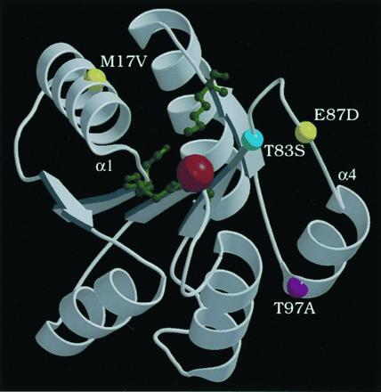
Figure 3.

Ribbon diagram of Mg2+-bound CheY crystal structure from coordinates (27), drawn with molscript (28) and raster3d (29). CheY active site residues D12, D13, D57, and K109 are depicted as green ball-and-stick forms; a red ball shows Mg2+. The locations of the equivalent PhoB residues are shown as colored balls: T83 (cyan), M17 and E87 (yellow), and T97 (purple). The amino termini of helix 1 and helix 4 are labeled α1 and α4, respectively.
