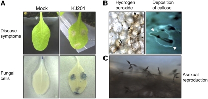
Figure 3.
Cellular responses of Arabidopsis to KJ201 infection. A, Three 5-μL drops of a fungal spore suspension (5 × 10⁵ spores mL−1) were placed on the leaves of 28- to 30-d-old Col-0 plants. Control plants were inoculated with distilled water (Mock). Leaves at 3 dpi are shown (top row). The same leaves were stained with trypan blue and observed with a microscope (bottom row). B, H2O2 accumulation was detected in Col-0 leaves inoculated with KJ201 using DAB staining (left). The appressoria are denoted as A. Aniline blue fluorochrome staining of the leaves revealed callose accumulation (arrowheads) around the penetrated sites after 24 h (right). C, Asexual reproduction of M. oryzae on infected Col-0 leaves.

