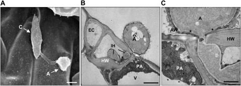
Figure 4.
Electron micrographs of Col-0 plants inoculated with M. oryzae strain KJ201. Plants inoculated with KJ201 (two 5-μL drops of a spore suspension at 5 × 10⁵ spores mL−1 in distilled water) were observed using scanning and transmission electron microscopy at 2 dpi. A, Scanning electron micrograph showing germinated conidia (C) forming an appressorium (A) on the leaf surface. Bar = 10 μm. B, Transmission electron micrograph showing infectious hyphae (IH) penetrating an epidermal cell (EC). The host cell wall (HW), papilla-like structure (PA), and vacuole (V) are indicated. Bar = 3 μm. C, Site of initial penetration, showing part of an appressorium and IP. The outer layer of the appressorial wall (AW) is more electron dense than the inner layer; a third wall layer (marked with asterisks) forms a thickened ring around the penetration pore, continuous with the IP wall. Bar = 1 μm.

