Figure 1.
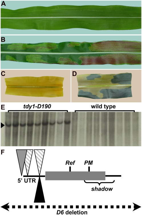
The cloning of Tdy1. A and B, Photographs of mature leaves. C and D, Photographs of segments from the same leaves, cleared and starch stained. A and C, Wild type. B and D, tdy1. E, Southern blot of tdy1-D190 mutants and wild-type siblings DNA probed with Mu1 (arrowhead). F, Schematic of mutations in different tdy1 alleles. Lines represent 5′ and 3′ untranslated regions (UTRs), the shaded box represents the Tdy1 protein-coding region, inverted triangles represent Mu insertions (striped, Mu1; gray, Mu8; black, Mu3), vertical lines indicate the locations of Ref and PM mutations, the bracket shows the region deleted in the shadow allele, and the dotted line indicates that the entire gene is deleted in the D6 allele. [See online article for color version of this figure.]

