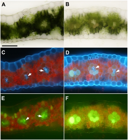
Figure 7.
CF movement along the symplastic pathway is retarded in tdy1 leaves. A, C, and E, Wild type. B, D, and F, tdy1-R chlorotic region. A and B, Transverse sections of mature leaf blades under bright field. C and D, Same sections under UV illumination. E and F, Same sections under blue light to image CF. Arrows indicate VP cells. Bar = 100 μm. [See online article for color version of this figure.]

