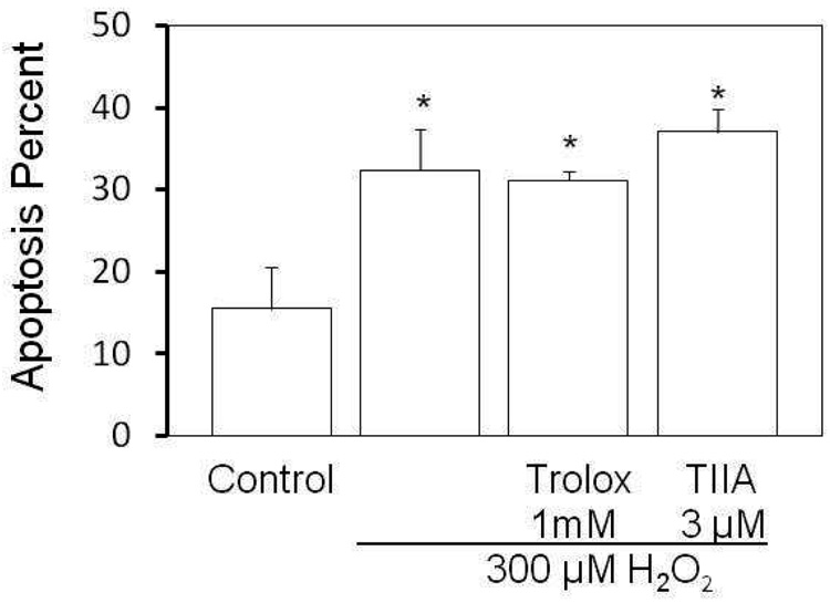
Figure 3.
Mitochondrial potential is not altered by TIIA. H2O2-induced apoptosis in J774 cells was analyzed by flow cytometry. J774 cells receiving vehicle, 1mM Trolox or 3µM TIIA pretreatment as in Figure 2 were incubated with 300 µM H2O2 for 4 hours. The mitochondrial membrane potential was assessed using the JC-1 dye (as described in Methods). Cells emitting green fluorescence were considered apoptotic. Five thousands cells were counted in one sample. The apoptosis percentage was calculated as: cells emitting green fluorescence/ total cells, presented as mean ± SEM and n = 3 in each group; *p<0.006 versus control.

