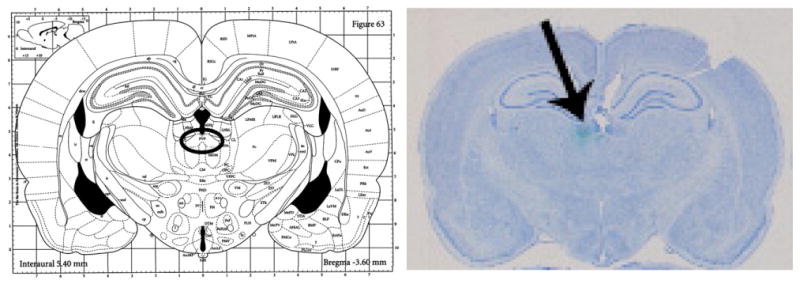
Fig. 1.

Bilateral guide cannula placement. Left, figure from Paxinos and Watson, 1986; the circle indicates the target area of the THPVP. Right, actual bilateral guide cannula placement. Cresyl violet stained coronal section showing cannula track (black arrow) and cannula end (green dye) marking the target area of the THPVP. (The specific placement coordinates for this micrograph correspond to −3.3 mm AP from bregma; ±0.2 mm ML from midline; −5.4 mm DV from skull.)
